
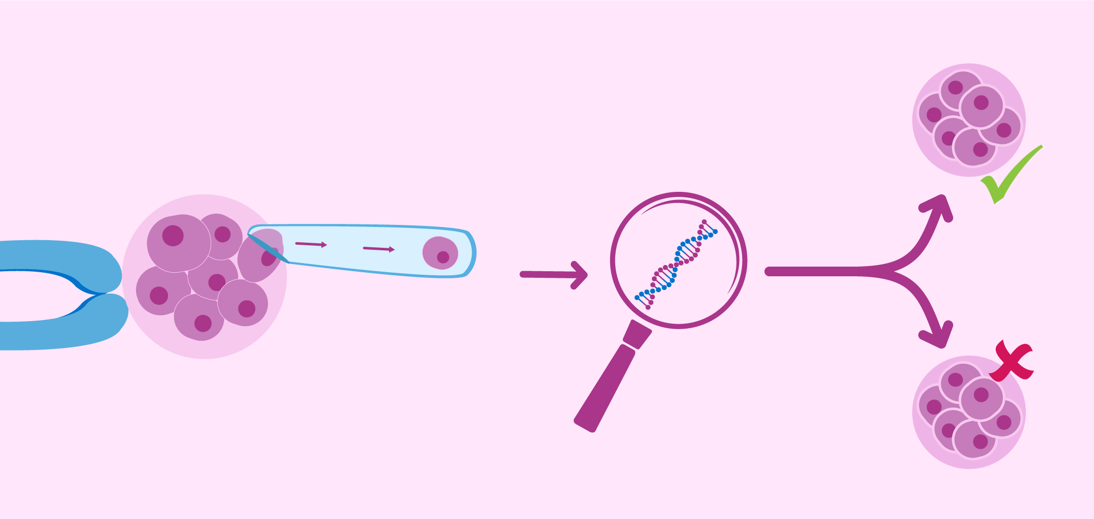
The researchers indicate that the preimplantation genetic diagnostic procedure is performed on the fifth day after fertilization, when the embryo reaches the state of “blastocyst” (day 5-6 after fertilization), which has the form of a vesicle delimited by a layer of outer cells, called trophoblast, and a mass of internal cells called embryoblast.
The trophoblast will be part of the future placenta, while the embryoblast will produce the embryo’s own body. Preimplantation genetic diagnosis (PGD) may not be effective in preventing premature spontaneous abortions, according to the results of a study conducted by a group of Italian and American researchers and doctors.
The samples are based on a considerable number of embryos frozen in blastocyst status, diagnosed as “chromosomally normal” by PGD and transferred almost the same amount to women, and a larger number of embryos not analyzed by PGD and transferred in women. According to the results, there are no notable differences between the frequency of spontaneous abortions between both groups.
The study, conducted with relatively high numbers of embryos, adds to other observations that confirm the doubts about the efficacy of PGD to detect chromosomal abnormalities of the future fetus. In this framework of ideas, a group of American researchers have developed a mathematical model, based on the distribution of cells in human embryos in the blastocyst state, and have concluded that it is necessary to analyze at least 27 cells of the trophoblast so that the margin of error is acceptable, something that would be hardly feasible with the survival of the embryo studied.
Take care of your health with the new digital medical center Pharmamedic.