
[et_pb_section bb_built=”1″][et_pb_row][et_pb_column type=”4_4″][et_pb_text _builder_version=”3.13.1″]
The clinical diagnosis of diseases such as cancer is mainly determined by the clinical suspicion, the physical examination and the last word of the specialist that establishes and confirms the presence of cancer cells in a tissue sample or biopsy.
Liquid biopsy represents a significant change, because it is a tool that can be performed many times during treatment and that has the ability to identify biomarkers in blood.
The liquid biopsy consists of extracting samples of blood, plasma, serum or urine to identify genetic variants and mutations. This procedure is much less invasive than the usual biopsy, which requires operation or puncture.
The system is in a phase prior to its application for the diagnosis of cancer, focused on monitoring the progression of the disease or the detection of genetic mutations in tumors that could suggest which drug should be used for its treatment.
The advances have been concentrated especially in the field of oncology, since it allows obtaining information when applying targeted therapies. The technique is especially interesting in the monitoring of cancer because during the course of treatment and the progression of the disease new genetic mutations are generated and multiple biopsies are usually necessary for decision making.
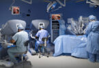
A new technology that facilitates the taking of breast tissue biopsies has been presented by the Center of Pathology of the Mama-Fundación Tejerina, in Spain, with the purpose of analyzing small lesions found in mammograms, in a faster and more energetic way.
The device is constituted by a selenium detector and Tomosynthesis technology, it works by making cutting planes of the breast.
The coupling of the table and the software make biopsies much more accurate, in addition, analyzes can be performed in shorter lapses, than with traditional biopsies, to allow microcalcifications or deformation in mammography.
This image and analysis tool is developed through a team and a work table that complements other available imaging methods, and allows the localization of microcalcification or distortions observed in mammography.
These and other innovations are now possible in Pharmamedic.
[/et_pb_text][/et_pb_column][/et_pb_row][/et_pb_section]