
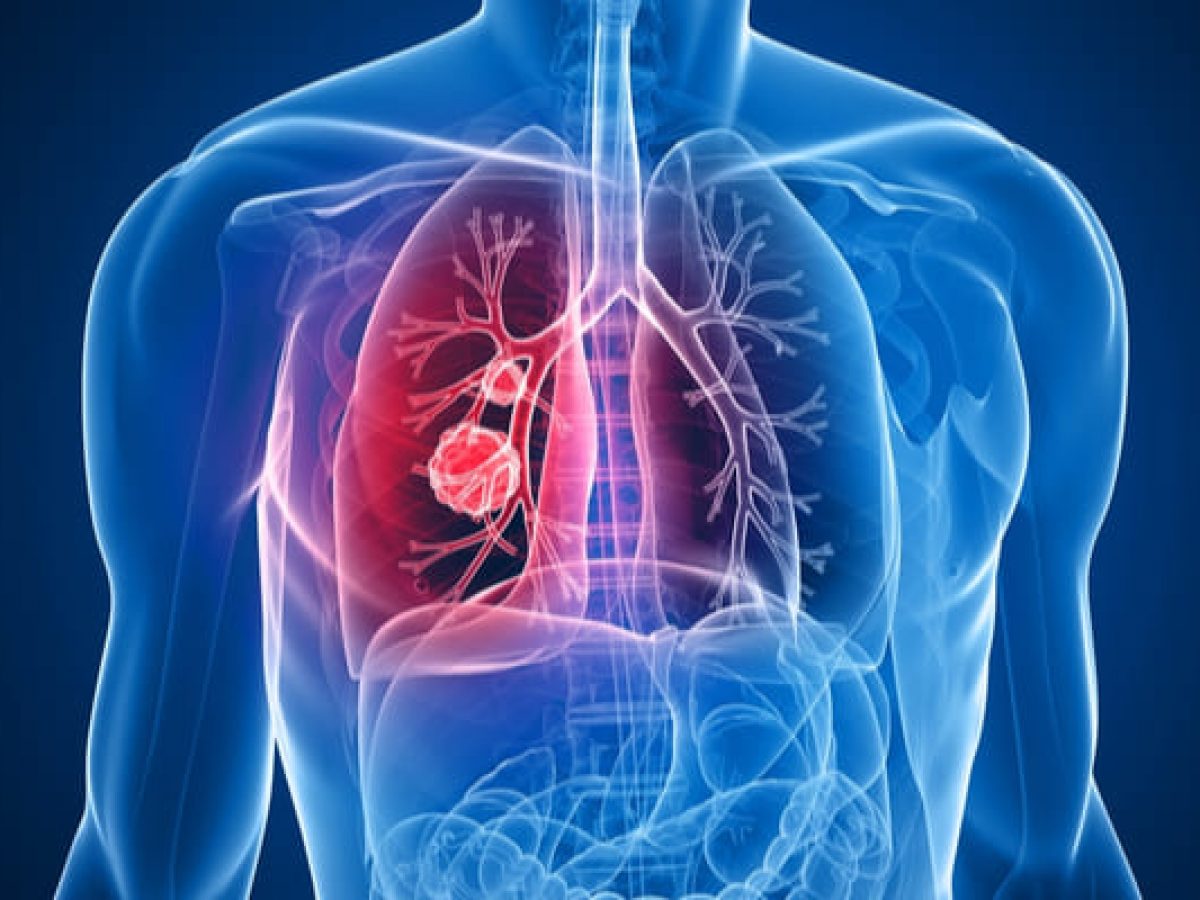
Pulmonary thromboembolism (PTE) is a blockage of an artery in the lungs by a substance that has moved from another part of the body through the bloodstream (thrombus).
Symptoms of a PE may include shortness of breath (dyspnea), chest pain, especially when inhaling, and coughing up blood (haemoptysis). Symptoms of a blood clot in the leg may also occur, such as a red, warm, swollen, and painful leg. Signs of PE include low blood oxygen levels, rapid breathing (tachypnea), rapid heart rate (tachycardia), and sometimes a mild fever. Severe cases can cause fainting, abnormally low blood pressure (hypotension), obstructive shock, and sudden death.
PE is usually the result of a blood clot in the leg that reaches the lung.
The risk of blood clots is increased with cancer, prolonged bed rest, smoking, stroke, certain genetic conditions, estrogen-based medications, pregnancy, obesity, and after some types of surgery. A small proportion of cases are due to embolization of air, fat, or amniotic fluid. Diagnosis is based on signs and symptoms in combination with test results. If the risk is low, a blood test known as D-dimer can rule out the condition. Otherwise, a CT with pulmonary angiography, pulmonary ventilation/perfusion scan, or Doppler ultrasound of the legs can confirm the diagnosis. Together, deep vein thrombosis and PE are known as venous thromboembolism (VTE).
On physical examination, the lungs are usually normal. Occasionally, a pleural friction rub can be heard over the affected area of the lung (mainly in PTE with infarction). Sometimes a pleural effusion is present that is exudative, detectable by decreased percussion note, audible breath sounds, and vocal resonance. Right ventricular tension can be detected as a left parasternal rise, a loud pulmonary component of the second heart sound, and/or increased jugular venous pressure. There may be a low-grade fever, particularly if there is associated pulmonary hemorrhage or infarction.
Because smaller pulmonary emboli tend to lodge in more peripheral areas without collateral circulation, they are more likely to cause pulmonary infarction and small effusions (both painful), but not hypoxia, dyspnea, or hemodynamic instability such as tachycardia.
Causes
After a first episode of PE, the search for secondary causes is usually brief. Only when a second PE occurs, and especially when this happens while still on anticoagulation therapy, is a further search for underlying conditions undertaken. This will include tests (“thrombophilia screening”) for factor V Leiden mutation, antiphospholipid antibodies, protein C and S, and antithrombin levels, and later prothrombin mutation, MTHFR mutation, factor VIII level, and hereditary coagulation abnormalities rarer.
Diagnosis
To diagnose a pulmonary embolism, a review of clinical criteria is recommended to determine the need for testing. In those at low risk, they are younger than 50 years old, have a heart rate less than 100 beats per minute, an oxygen level greater than 94% on room air, and no leg swelling, coughing up blood, surgery, or trauma to the body. past four weeks, previous blood clots, or estrogen use, no further testing is usually needed.