[et_pb_section bb_built=”1″][et_pb_row][et_pb_column type=”1_2″][et_pb_text _builder_version=”3.13.1″]
Bronchoscopy, also known as fibrobronchoscopy, is a medical test used to diagnose and treat diseases of the airways and lungs.
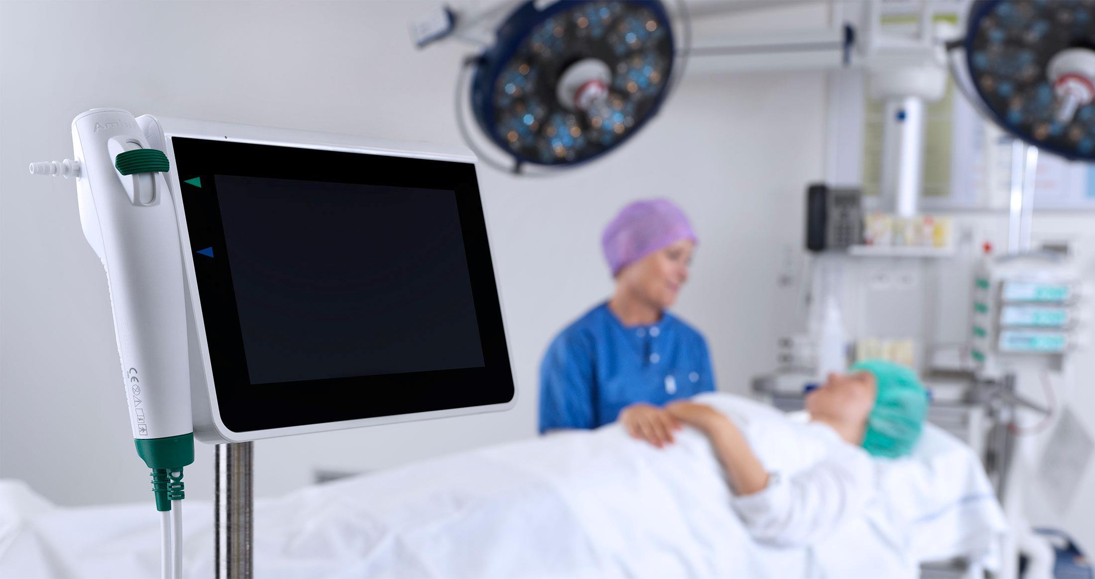
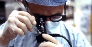
It is performed by means of a bronchoscope, an apparatus that consists of a tube about half a centimeter in diameter and of very variable length, depending on the age of the patient, who has a video camera at its end. It allows to clearly see the inside of the airways on a monitor. The tube of the bronchoscope also has several channels in its interior, where different instruments can be inserted, which allow to perform diagnostic tests such as taking biopsies, or even perform treatments such as the cauterization of bleeding vessels or the removal of polyps. .
There are two types of bronchoscopes: The flexible bronchoscope is the most commonly used. It consists of a flexible and thin tube that is inserted through the nose and allows to reach very far in the airway with greater comfort for the patient. No general anesthesia is required to perform the test. The rigid bronchoscope consists of a thicker, shorter and stiffer tube. It is inserted through the mouth and can not reach as far as the flexible bronchoscope. For its use it is required that the patient is under a general anesthesia. The advantage of the rigid bronchoscope is that its light is very thick. It is used in cases of massive hemorrhages that can prevent a correct visibility with the flexible bronchoscope. It is also used to perform large biopsies or to remove large foreign bodies that can not be removed with the flexible bronchoscope. There are procedures such as the dilatation of a bronchus that has been narrowed, or the use of the laser to destroy a tumor, which are performed by means of the rigid bronchoscope.
The most frequent reasons for which you can request these tests are: Identify the cause of some symptoms such as chronic cough, haemoptysis or difficulty breathing. These bronchoscopes are used to take samples or biopsies of the respiratory tract, the lung, or nearby lymph nodes, to study possible respiratory infections, tumors, or other lung diseases. Similarly, to diagnose and evaluate the extent of lung cancer, extract foreign bodies from the airway, treat bleeding that occurs in the airways, dilate areas of the airway that may have been narrowed by a tumor or other pathologies, allows the placement of a stent, which is like a self-expanding spring, which dilates the stenotic zone and keeps it open, treating lung cancer by means of the laser or placing radioactive substances nearby (brachytherapy).
[/et_pb_text][/et_pb_column][et_pb_column type=”1_2″][et_pb_text _builder_version=”3.13.1″]
Chronic diseases of the lower respiratory tract. Some of the most common diseases in the field of pulmonology are classified in this group, among them chronic bronchitis, chronic obstructive pulmonary disease, pulmonary emphysema, asthma and bronchiectasis.
Each year about 3 million people undergo a lung biopsy. When possible, specialists use bronchoscopes to perform it, a tiny device that carries a small camera and small surgical tools. This system allows minimal risk and trauma for the patient. However, many times the tumor on which the biopsy must be performed is located in the deepest part of the lung, in a labyrinth of respiratory conducts and it is impossible to reach it with the bronchoscope. The alternative solution is a surgical intervention through an incision in the chest, an intervention that increases the risk of a lung collapse, becoming infected or even dying the patient.
But one of the latest technological advances in the field of medicine is a new process that allows the bronchoscope to enter the depths of the lung. The system starts with existing technology that creates images of the lung in 3-D from CT-type ultrasounds.
The novelty of this new invention is that it is possible to obtain data on the situation of the bronchoscope inside the lung, and then contrast them with the 3-D image. A sensor at the tip of the bronchoscope reports its situation to an antenna on a table that is placed under the patient.
Then a program transfers the exact location of the bronchoscope to the virtual image in 3 dimensions, allowing the surgeon to see exactly where the bronchoscope is and guide it through the smaller airways to the tumor and avoiding the need to resort to a surgical intervention to perform the biopsy. The projection for this novel invention is that it will avoid almost one million cases of lung surgery per year.
These and other innovations are also possible in Pharmamedic.
[/et_pb_text][/et_pb_column][/et_pb_row][/et_pb_section]